45 microscope images with labels
Parts of Microscope, Function, Names & Labeled Diagram - slidingmotion Microscope parts labeled diagram gives us all the information about its parts and their position in the microscope. Microscope Parts Labeled Diagram The principle of the Microscope gives you an exact reason to use it. It works on the 3 principles. Magnification Resolving Power Numerical Aperture. Parts of Microscope Head Base Arm Eyepiece Lens Microscope Types (with labeled diagrams) and Functions This is an advanced microscope that has specific application in viewing, observing and measuring the optical thickness and phase of completely transparent specimens and objects. A tiny interferometer is used and a specimen is placed on beam path of it. This path is split and then rejoined to create two superimposed images of the specimen in focus.
Microscope Drawing And Label - Painting Valley Are you looking for the best images of Microscope Drawing And Label? Here you are! We collected 33+ Microscope Drawing And Label paintings in our online museum of paintings - PaintingValley.com. ADVERTISEMENT LIMITED OFFER: Get 10 free Shutterstock images - PICK10FREE label microscope diagram compound parts light labeling functions microscopic

Microscope images with labels
Microscope Labeling - The Biology Corner The google slides shown below have the same microscope image with the labels for students to copy. I often spend the first day walking students through the steps and having them look at a single slide as we do the steps. Students are often very enthusiastic about using microscopes and will try to start with the high power objective. Microscope Labeling Game - PurposeGames.com About this Quiz. This is an online quiz called Microscope Labeling Game. There is a printable worksheet available for download here so you can take the quiz with pen and paper. This quiz has tags. Click on the tags below to find other quizzes on the same subject. Science. Microscope Labeled Pictures, Images and Stock Photos Browse 48 microscope labeled stock photos and images available, or start a new search to explore more stock photos and images. Newest results Fluorescent Imaging immunofluorescence of cancer cells growing... Plant Tissue Systems vector illustration. Labeled biology... Microscope diagram vector illustration. Labeled zoom instrument...
Microscope images with labels. Microscope Parts and Functions With Labeled Diagram and Functions How ... Coarse adjustment: Brings the specimen into general focus. Fine adjustment: Fine tunes the focus and increases the detail of the specimen. Nosepiece: A rotating turret that houses the objective lenses. The viewer spins the nosepiece to select different objective lenses. Objective lenses: One of the most important parts of a compound microscope ... 300+ Free Microscope & Laboratory Images - Pixabay Upload 399 Free images of Microscope Related Images: laboratory science bacteria research scientist lab biology chemistry medical Find your perfect microscope image. Free pictures to download and use in your next project. 399 Free images of Microscope / 4‹ › 26+ Picture Of A Microscope With Label PNG - Berita Seputar Dunia ... 26+ Picture Of A Microscope With Label PNG. Microscopes are specially created to magnify the image of the subject being studied. Students label the parts of the microscope in this photo of a basic laboratory light microscope. Microscope Drawing And Label at GetDrawings | Free download from getdrawings.com. Sharper microscope images wanted: labels need not apply - Ars Technica Sharper microscope images wanted: labels need not apply 9 posts JournalBot. Ars Legatus Legionis et Subscriptor. Registered: Apr 5, 2005. Posts: 101520. Posted: Thu Oct 30, 2008 2:06 pm ...
PDF Parts of a Microscope Printables - Homeschool Creations Label the parts of the microscope. You can use the word bank below to fill in the blanks or cut and paste the words at the bottom. Microscope Created by Jolanthe @ HomeschoolCreations.net. Parts of a eyepiece arm stageclips nosepiece focusing knobs illuminator stage objective lenses Compound Microscope Parts - Labeled Diagram and their Functions - Rs ... The eyepiece (or ocular lens) is the lens part at the top of a microscope that the viewer looks through. The standard eyepiece has a magnification of 10x. You may exchange with an optional eyepiece ranging from 5x - 30x. [In this figure] The structure inside an eyepiece. The current design of the eyepiece is no longer a single convex lens. Parts of the Microscope with Labeling (also Free Printouts) Microscopes are specially created to magnify the image of the subject being studied. This exercise is created to be used in homes and schools. the microscope layout, including the blank and answered versions are available as pdf downloads. Click to Download : Label the Parts of the Microscope (A4) PDF print version. Microscope picture label Flashcards | Quizlet Start studying Microscope picture label. Learn vocabulary, terms, and more with flashcards, games, and other study tools.
8,516 Microscope Slide Label Templates - TemplateMonster Minimalist is a beautiful PowerPoint template you can use to design professional presentations for business and agency meetings and events. The template comes with 140 unique slides in HD resolution and features image placeholders, world maps, icons, and much more.Key Features : 140 different slides, Simple. Parts of a Simple Microscope - Labeled (with diagrams) image 2: A simple microscope commonly used by students for studying minute objects. image source: imimg.com. picture 3: It is the latest design of a simple microscope - advanced features than the conventional simple microscopes. ... image 5: A modern simple microscope with the different parts labeled. image source: laboratoryinfo.com. The ... Microscope Labeling Practice Quiz Practice labeling a compound microscope. Do you know all the parts? Practice labeling a compound microscope. Do you know all the parts? English en. Login. Login Register ... North America - Countries, Waterways & Neighbors 22p Image Quiz. Genetics Vocabulary Practice 26p Text Game. Neuron Diagram 7p Image Quiz. PurposeGames Create. Play. Learn ... Simple Microscope - Diagram (Parts labelled), Principle, Formula and Uses A simple microscope consists of Optical parts Mechanical parts Labeled Diagram of simple microscope parts Optical parts The optical parts of a simple microscope include Lens Mirror Eyepiece Lens A simple microscope uses biconvex lens to magnify the image of a specimen under focus.
Microscope Picture To Label - Micropedia
Labeling the Parts of the Microscope | Microscope World Resources Labeling the Parts of the Microscope This activity has been designed for use in homes and schools. Each microscope layout (both blank and the version with answers) are available as PDF downloads. You can view a more in-depth review of each part of the microscope here. Download the Label the Parts of the Microscope PDF printable version here.

Microscope With Labels free vector | Download it now!
Microscope Images Labeled | Virtual Anatomy Lab VAL - ncccval Microscope Images Labeled | Virtual Anatomy Lab VAL
Search in gallery
Parts of a Compound Microscope - Labeled (with diagrams) It is used to carry the microscope and at the same time connect the base of the microscope to the head. (1, 2, 3, and 4) Image 3: A compound microscope with a corresponding label of the different parts.

Haversian System
Microscope Imaging Station. Gallery. - Exploratorium Chloroplasts and mitochondria move within Elodea leaf cells; nuclei are also visible as clear, fried-egg-shaped structures. Elodea are common freshwater aquarium plants. An elodea leaf was mounted in pondwater between a slide and coverslip with a silicon spacer. Images were taken on an inverted compound microscope using a 40x DIC objective and ...

Microscopic Structure of Bone | ClipArt ETC
Parts of a microscope with functions and labeled diagram - Microbe Notes Optical parts of a microscope and their functions The optical parts of the microscope are used to view, magnify, and produce an image from a specimen placed on a slide. These parts include: Eyepiece - also known as the ocular. This is the part used to look through the microscope. Its found at the top of the microscope.

31 Picture Of A Microscope To Label - Labels For Your Ideas
Parathyroid Gland Histology with Microscope Slide Image and Labeled ... Parathyroid Gland Histology with Microscope Slide Image and Labeled Diagram 09/12/2021 07/12/2021 by anatomylearner There are two or more pairs of parathyroid glands located on the posterior surface of the thyroid gland. You will find two main types of cells (chief and oxyphils) in the parathyroid gland histology slide.
Label a microscope - Teaching resources
Compound Microscope with labels Stock Vector | Adobe Stock Download Compound Microscope with labels Stock Vector and explore similar vectors at Adobe Stock. Adobe Stock. Photos Illustrations Vectors Videos Audio Templates Free Premium Editorial Fonts. ... Get 10 free Adobe Stock images. Start now. Get 10 free images. Unlock 200M+ assets in our full collection.
Search in gallery
Microscope With Labeled Parts And Functions Microscope is a revolutionized scientific instrument which is used in research laboratories to examine the small objects that are not clearly visible and can't be seen by the naked eye. They are derived from Ancient Greek words "mikrós skopeîn" as mikrós means "small" and skopeîn mean "to look" or "see".

Lysosomes Dr.Jastrow's electron microscopic atlas
PDF Label parts of the Microscope Label parts of the Microscope: . Created Date: 20150715115425Z
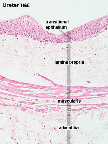
ANAT2511 Urinary System - Embryology
Skin Histology Slide Identification - Best Place to Learn Veterinary ... I would like to show you the different histological features from both thick and thin skin histology slides with a labeled diagram. I hope these skin microscope slide labeled diagrams might help you to identify and learn all the structures. If you need more skin microscope slide labeled diagram, please follow anatomy learner on social media. I ...

labels of a compound microscope microscope boxed - Top Label Maker
Label the microscope — Science Learning Hub Use this interactive to identify and label the main parts of a microscope. Drag and drop the text labels onto the microscope diagram. eye piece lens: The lens you look through - normally 10x or 15x magnification. eye piece lens. coarse focus adjustment: Moves the lens up or down and adjusts focus. coarse focus adjustment.

Microscope Journal – The Indigo Teacher
Microscope Labeled Pictures, Images and Stock Photos Browse 48 microscope labeled stock photos and images available, or start a new search to explore more stock photos and images. Newest results Fluorescent Imaging immunofluorescence of cancer cells growing... Plant Tissue Systems vector illustration. Labeled biology... Microscope diagram vector illustration. Labeled zoom instrument...

Labeling The Microscope - PurposeGames
Microscope Labeling Game - PurposeGames.com About this Quiz. This is an online quiz called Microscope Labeling Game. There is a printable worksheet available for download here so you can take the quiz with pen and paper. This quiz has tags. Click on the tags below to find other quizzes on the same subject. Science.
Quia - Protist Vocabulary
Microscope Labeling - The Biology Corner The google slides shown below have the same microscope image with the labels for students to copy. I often spend the first day walking students through the steps and having them look at a single slide as we do the steps. Students are often very enthusiastic about using microscopes and will try to start with the high power objective.

Label the microscope Quiz

Labeling a Compound Microscope - PurposeGames
Zoeken in galerij
Post a Comment for "45 microscope images with labels"